

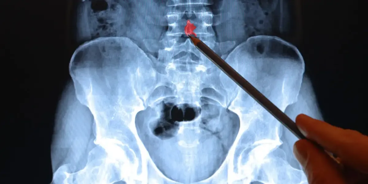
Imaging has become a central part of how spine conditions are evaluated, often shaping how patients understand their diagnosis. Seeing a scan can make a condition feel more concrete, especially when symptoms have been difficult to explain. At the same time, imaging does not always provide a complete or straightforward answer. Dr. Larry Davidson, a board-certified neurosurgeon with fellowship training in complex spinal surgery, has seen how patients sometimes place too much weight on what appears in an MRI or CT scan without fully understanding how those findings relate to their symptoms.
Understanding the role of imaging requires a broader perspective. These tools are designed to reveal structural details, but they do not measure pain, function or how the body responds to movement. Interpreting imaging correctly depends on how well those findings align with the patient’s overall clinical picture.
How Imaging Fits Into the Diagnostic Process
Imaging is typically used to support, not replace, a clinical evaluation. When patients present with persistent pain or neurological symptoms, physicians begin by assessing how those symptoms developed and how they behave over time. This initial step often provides important clues about what may be causing the issue.
When imaging is introduced, it serves as a way to confirm or refine those initial impressions. MRI scans are commonly used to evaluate soft tissues such as discs, nerves and ligaments, while CT scans provide more detailed information about bone structures. Each modality offers a different perspective, and together they can help create a more complete view of the spine.
What MRI and CT Scans Can Reveal
MRI is particularly effective at identifying conditions that affect soft tissue structures. Disc herniations, nerve compression and changes in the spinal cord are often clearly visible, allowing physicians to identify areas that may be contributing to symptoms.
CT scans, on the other hand, are better suited for evaluating bone-related issues. They can reveal fractures, bone spurs and alignment changes that may not be as easily detected with MRI. In some cases, both types of imaging are used together to provide a more detailed understanding of the condition. These tools offer valuable insight into the anatomy of the spine. However, they are limited to showing structure, not function. They cannot fully explain how a patient experiences pain or how symptoms affect daily activity.
When Imaging Findings Do Not Match Symptoms
One of the more challenging aspects of spine diagnosis is that imaging findings do not always align with what a patient feels. It is common for individuals without pain to show signs of disc degeneration or minor abnormalities on MRI. At the same time, patients with significant discomfort may have imaging results that appear relatively unremarkable.
A disconnect can be confusing. Patients may wonder why they feel pain when nothing significant appears on a scan, or why abnormalities are present without corresponding symptoms. These situations highlight the complexity of spine conditions and the role of the nervous system in shaping how pain is perceived. Because of this, imaging must be interpreted carefully. Findings are only meaningful when they correlate with the patient’s symptoms and clinical presentation.
Why Correlation Is Critical
Accurate diagnosis depends on correlating imaging results with clinical findings. It means determining whether a visible abnormality is actually responsible for the symptoms being experienced. Without this step, there is a risk of focusing on findings that may not be clinically relevant.
For example, a disc bulge may appear on imaging, but if it does not correspond to the patient’s pain pattern or neurological symptoms, it may not be the source of the issue. Conversely, a smaller or less obvious finding may be highly significant if it aligns closely with the patient’s experience. This process requires careful evaluation and often relies on both clinical expertise and patient input.
When Imaging Becomes More Important
While imaging is not always necessary in the early stages of back or neck pain, it becomes more relevant when symptoms persist or change. Ongoing pain that does not respond to conservative care often prompts further investigation to find what may be contributing to the condition.
Imaging is also important when neurological symptoms are present. Numbness, tingling or weakness may indicate nerve involvement, which often requires visualization to determine the cause. In these cases, imaging helps identify whether structural changes are affecting the nervous system. The timing of imaging matters. Using it too early may not provide useful information, while delaying it in the presence of concerning symptoms can limit the ability to address the issue effectively.
Avoiding Overinterpretation of Imaging Results
One of the challenges in modern spine care is avoiding overinterpretation of imaging findings. Because scans can reveal abnormalities even in individuals without symptoms, there is a risk of attributing pain to findings that may not be clinically relevant.
It can lead to unnecessary treatments or interventions that do not address the actual source of discomfort. It can also create anxiety for patients who may interpret any abnormal finding as a serious issue. A balanced approach involves viewing imaging as one piece of the diagnostic process. Physicians focus on how findings fit within the broader clinical picture, rather than treating the image alone.
How Imaging Fits Into the Bigger Clinical Picture
Spine health cannot be understood through imaging alone. A combination of structural, neurological and behavioral factors influences pain, movement and function. While imaging provides valuable insight into anatomy, it does not capture the full experience of the patient.
Dr. Larry Davidson highlights the importance of aligning imaging findings with the patient’s symptoms before making treatment decisions, particularly when considering more advanced interventions. This approach reflects a broader view of diagnosis, one that prioritizes meaningful connections over isolated findings. By understanding both the value and the limitations of imaging, patients can approach their diagnosis with greater clarity. Instead of relying solely on what a scan shows, they can consider how those findings fit into the larger picture of their condition and their overall care.
How Imaging Guides Treatment Decisions
When used appropriately, imaging plays an important role in guiding treatment. It helps confirm diagnoses, identify the location of structural issues, and support decisions about both non-surgical and surgical care.
For non-surgical treatment, imaging can help determine which areas of the spine should be the focus of therapy. For surgical planning, it provides detailed information that allows for more precise intervention, reducing unnecessary disruption to surrounding structures. This level of detail supports more individualized care. Treatment plans can be tailored to the specific characteristics of the condition rather than based on general assumptions.